
Global Medical Services
International Medical Center
 Diseases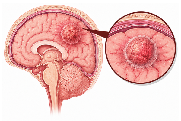
Brain metastases are cancer cells that spread to the brain from primary tumors elsewhere. Learn about causes, symptoms, treatment options, and prognosis at Guangzhou Fosun Chancheng Hospital.
Brain metastases occur when cancer cells spread hematogenously to the brain parenchyma. They are the most common intracranial tumors in adults (10x more common than primary brain tumors):
Primary Sources: Lung cancer (40-50%), breast cancer (15-25%), melanoma (10-15%), renal cell carcinoma (5-10%), colorectal cancer (3-5%). Less common: thyroid, gynecologic, germ cell tumors. Unknown primary in 5-10%.
Mechanism: Hematogenous dissemination via arterial circulation. Tumor cells arrest at gray-white matter junction (narrow vessel caliber). Extravasation and colonization dependent on tumor-host microenvironment interactions (seed and soil hypothesis).
Molecular Biology: Tumor cells must cross blood-brain barrier (BBB). Role of adhesion molecules, matrix metalloproteinases, angiogenesis (VEGF), and evasion of CNS immune surveillance. Specific gene signatures associated with CNS tropism (e.g., HER2, EGFR mutations in lung cancer, triple-negative/basal subtype in breast cancer).
Distribution: Cerebral hemispheres 80% (frontal > parietal > temporal > occipital), cerebellum 15%, brainstem 5%. Matches regional cerebral blood flow.
Headache: Present in 40-50%. Often dull, constant, worse in morning or with Valsalva. May be localized or generalized. Papilledema in 15-25%.
Focal Neurological Deficits: Present in 30-40%. Hemiparesis (frontal/parietal), visual field deficits (occipital), aphasia (dominant temporal/frontal), sensory deficits, gait ataxia (cerebellar).
Seizures: Presenting symptom in 15-20%. Focal onset with or without secondary generalization. More common with melanoma and hemorrhagic metastases.
Cognitive Changes: Memory impairment, personality changes, executive dysfunction, confusion, lethargy. May be subtle and gradual.
Nausea/Vomiting: From increased intracranial pressure. Often worse in morning.
Stroke-like Presentation: Sudden onset from hemorrhage into metastasis (melanoma, RCC, choriocarcinoma are highly hemorrhagic).
Stereotactic Radiosurgery (SRS): First-line for limited metastases (1-4 lesions). Delivers highly conformal radiation in single fraction (15-24 Gy). Local control 80-90%. Cognitive preservation compared to WBRT. Can be repeated for new metastases.
Whole Brain Radiotherapy (WBRT): For multiple (>10) brain metastases, poor performance status, or rapid progression. 30 Gy in 10 fractions standard. Hippocampal avoidance WBRT + memantine reduces cognitive toxicity.
Surgery: Indicated for single large metastasis (>3cm) with mass effect, diagnostic uncertainty, or controlled systemic disease. Gross total resection + SRS to cavity improves local control vs. SRS alone.
Systemic Therapy: Targeted agents with CNS penetration: osimertinib (EGFR-mutant NSCLC), alectinib/lorlatinib (ALK-rearranged NSCLC), tucatinib + trastuzumab + capecitabine (HER2+ breast cancer), immunotherapy (ipilimumab + nivolumab for melanoma).
Corticosteroids: Dexamethasone 4-8 mg/day for symptomatic peritumoral edema. Taper as soon as clinically feasible.
Graded Prognostic Assessment (GPA): Based on age, KPS, number of metastases, extracranial disease status. Median survival by GPA: 3-6 months (poor), 6-12 months (intermediate), 12-24+ months (favorable).
Diagnosis-Specific Prognosis: Breast cancer: 10-15 months median. HER2+ with targeted therapy: 18-24+ months. Lung cancer: EGFR/ALK+ with targeted therapy: 18-24+ months. Melanoma: immunotherapy responders: 12-24+ months. RCC: 8-12 months. GI cancers: 3-6 months.
Prognostic Factors (favorable): Age <65>
Prognostic Factors (unfavorable): Age >65, KPS <70>
Screening: No routine brain screening for asymptomatic patients. MRI brain for any cancer patient with new neurological symptoms. Consider surveillance MRI for high-risk patients (stage III-IV NSCLC, melanoma stage III-IV, HER2+ breast cancer).
Seizure Prophylaxis: Not routinely recommended. Antiepileptic drugs only for patients who have had a seizure. Levetiracetam preferred (non-enzyme inducing, fewer drug interactions). Avoid phenytoin (CYP450 induction).
Steroid Management: Use lowest effective dexamethasone dose. Prophylactic PPI or H2 blocker for GI protection. Monitor blood glucose, watch for steroid myopathy, insomnia, psychiatric effects. Pneumocystis jirovecii pneumonia prophylaxis for prolonged high-dose steroids.
VTE Prophylaxis: High risk of venous thromboembolism. Consider prophylactic anticoagulation in hospitalized patients. LMWH preferred over warfarin for treatment.
Radiation Necrosis: Differentiate from tumor progression (MRI perfusion, MRS, PET). May occur 6-24 months post-SRS. Treated with steroids, bevacizumab, or surgical resection for refractory cases.
Concerned about Brain Metastases? Submit your medical records for a professional evaluation.
Upload Documents View Patient CasesPrepare pathology reports, imaging scans, genetic testing reports and prior treatment records for faster review.