
Global Medical Services
International Medical Center
 Diseases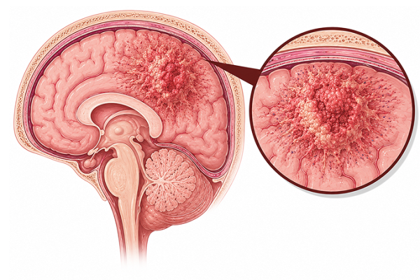
Brain glioma is a primary brain tumor arising from glial cells. Learn about its types, causes, symptoms, treatment, and prognosis at Guangzhou Fosun Chancheng Hospital.
Gliomas are primary CNS tumors arising from glial cells (astrocytes, oligodendrocytes, ependymal cells). They account for 30% of all brain tumors and 80% of malignant brain tumors:
Genetic Mutations: IDH1/IDH2 mutations (defining molecular marker in lower-grade gliomas, portend better prognosis). 1p/19q codeletion (diagnostic for oligodendroglioma, predicts chemosensitivity). MGMT promoter methylation (predicts temozolomide response, better prognosis). ATRX, TP53, TERT promoter mutations. EGFR amplification, PTEN deletion, CDKN2A/B deletion (glioblastoma). H3 K27M mutation (diffuse midline glioma).
Ionizing Radiation: Only established environmental risk factor. Therapeutic cranial radiation (e.g., for childhood leukemia, tinea capitis). No clear association with diagnostic radiation, EMF, or cell phones.
Genetic Syndromes: Neurofibromatosis type 1 (optic pathway glioma), NF2, Li-Fraumeni syndrome (TP53), Turcot syndrome, Lynch syndrome, tuberous sclerosis (SEGA), Ollier disease/Maffucci syndrome.
WHO Classification (2021): Adult-type diffuse gliomas: Astrocytoma IDH-mutant (grade 2-4), Oligodendroglioma IDH-mutant 1p/19q codeleted (grade 2-3), Glioblastoma IDH-wildtype (grade 4). No longer uses grade 1-4 alone; integrates histologic and molecular features.
Headache: Present in 50-60%. Dull, constant, worse in morning or with recumbency. From increased ICP or traction on pain-sensitive structures. Nausea/vomiting often accompany morning headaches.
Seizures: Presenting symptom in 30-50%. More common in low-grade gliomas involving cortex. Focal aware or focal with impaired awareness. May be first manifestation for years before diagnosis of low-grade gliomas.
Focal Neurological Deficits: Progressive hemiparesis, sensory loss, visual field defects, aphasia (depending on location). Frontal lobe: personality changes, disinhibition, executive dysfunction. Temporal lobe: memory, auditory, language. Parietal: sensory, spatial neglect. Occipital: visual.
Cognitive and Behavioral Changes: Memory impairment, confusion, personality changes, psychomotor slowing. May be subtle and attributed to depression or aging.
Increased ICP Symptoms: Papilledema, diplopia (CN VI palsy from stretching), nausea/vomiting, decreased consciousness (late).
Surgery: Maximal safe resection is standard. Extent of resection correlates with survival. Awake craniotomy with intraoperative mapping for eloquent cortex. Fluorescence-guided surgery (5-ALA) improves extent of resection for GBM. Re-resection for recurrent disease in selected patients.
Radiation Therapy: Focal fractionated RT 60 Gy in 30 fractions (grade 4), 54 Gy in 27 fractions (grade 3), 50-54 Gy (grade 2). Target: T2/FLAIR abnormality + 1-2 cm margin (grade 2-3) or enhancing tumor + margin (GBM). Hypofractionated RT for elderly/poor KPS: 40 Gy in 15 fractions.
Chemotherapy: Temozolomide: concurrent with RT (75 mg/m² daily) + adjuvant (150-200 mg/m² days 1-5 q28 x 6 cycles) for GBM (Stupp protocol). PCV (procarbazine, lomustine, vincristine) for 1p/19q codeleted oligodendroglioma. Lomustine for recurrent GBM.
Tumor Treating Fields (TTF): Optune device delivers alternating electric fields. Adjuvant for newly diagnosed GBM (EF-14 trial: improved OS 20.9 vs 16.0 months).
Targeted Therapy: Vorasidenib (IDH1/2 inhibitor) for IDH-mutant grade 2 glioma. BRAF/MEK inhibitors for BRAF V600E-mutant gliomas.
Glioblastoma IDH-wildtype (Grade 4): Median OS 14-16 months (Stupp protocol). 2-year survival 27%, 5-year survival 5-10%. Favorable: age <50>
Astrocytoma IDH-mutant (Grade 4): Median OS 31-48 months. Better prognosis than IDH-wildtype GBM.
Astrocytoma IDH-mutant (Grade 2-3): Median OS 5-10+ years. Grade 2: 10-15 years. Grade 3: 5-10 years.
Oligodendroglioma IDH-mutant 1p/19q codeleted (Grade 2-3): Best prognosis. Median OS 10-20+ years. Grade 3: 8-14 years with PCV + RT.
Prognostic Factors (favorable): IDH mutation, 1p/19q codeletion, MGMT promoter methylation, young age, high KPS, gross total resection, oligodendroglial histology, lower grade.
Prognostic Factors (unfavorable): IDH wildtype, unmethylated MGMT, age >60, low KPS, biopsy only, H3 K27M mutation (midline), CDKN2A/B deletion, EGFR amplification.
Seizure Management: Antiepileptic drugs for patients with seizures. Levetiracetam or lacosamide preferred (non-enzyme inducing). Avoid prophylactic AEDs in seizure-naive patients. Monitor for drug interactions (especially enzyme-inducing AEDs with chemotherapy).
Thromboembolism Prophylaxis: Glioma patients have highest VTE risk among all cancers (20-30%). Consider prophylactic LMWH in perioperative and hospitalized settings. Therapeutic anticoagulation for established VTE (LMWH preferred).
Corticosteroids: Dexamethasone for peritumoral edema. Lowest effective dose, shortest duration. Monitor glucose, blood pressure, myopathy. PJP prophylaxis for prolonged use. Taper gradually to avoid adrenal insufficiency.
Neurocognitive Monitoring: Baseline neuropsychological evaluation. Cognitive rehabilitation. Memantine during WBRT. Hippocampal-sparing RT techniques.
Pseudoprogression vs. True Progression: Common after chemoradiation (20-30%). Increased enhancement 3-6 months post-RT, then stabilizes/improves. MRI perfusion (rCBV), MRS, and PET help differentiate. RANO criteria for response assessment.
Supportive Care: Physical/occupational/speech therapy. Driving assessment (seizure risk). Psychological support for patients and caregivers. Palliative care early integration.
Concerned about Brain Glioma? Submit your medical records for a professional evaluation.
Upload Documents View Patient CasesPrepare pathology reports, imaging scans, genetic testing reports and prior treatment records for faster review.