
Global Medical Services
International Medical Center
 Diseases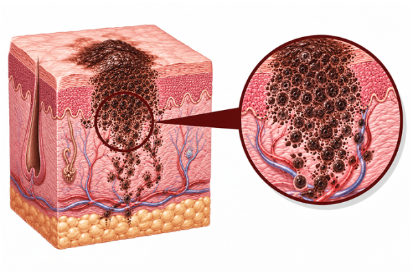
Melanoma is an aggressive form of skin cancer arising from melanocytes. Learn about its causes, types, symptoms, treatment, and prognosis at Guangzhou Fosun Chancheng Hospital.
Melanoma arises from malignant transformation of melanocytes (pigment-producing cells). Incidence rising faster than any other cancer worldwide:
UV Radiation: Most significant environmental risk factor. Intermittent intense sun exposure (blistering sunburns, especially childhood/adolescence) more strongly associated than cumulative exposure. UVA and UVB both mutagenic. Tanning bed use increases risk 75% if started before age 35.
Genetic Mutations: BRAF V600E/K mutations (40-60% of cutaneous melanomas), NRAS mutations (15-20%), NF1 mutations (10-15%), KIT mutations (acral/mucosal). CDKN2A (p16INK4a/p14ARF) germline mutations in familial melanoma. TERT promoter mutations (70%). PTEN loss.
Phenotypic Risk Factors: Fair skin (Fitzpatrick I-II), red/blonde hair, blue/green eyes, freckling, >50 melanocytic nevi, atypical/dysplastic nevi, giant congenital nevi (>20cm). Family history: 10% have first-degree relative with melanoma.
Subtypes: Superficial spreading (70%, intermittent sun), nodular (15-20%, rapid growth), lentigo maligna melanoma (5-15%, chronic sun), acral lentiginous (5-10%, palms/soles/nails, most common in dark-skinned), mucosal (<2>
ABCDE Criteria: Asymmetry (one half unlike the other), Border irregularity (notched, scalloped, poorly defined), Color variation (different shades of brown, black, red, white, blue), Diameter >6mm (pencil eraser size), Evolution (changing in size, shape, color, elevation, or new symptom: bleeding, itching, crusting).
Ugly Duckling Sign: A mole that looks different from all other moles on the individual. More sensitive than ABCDE alone. Patients often identify their own melanoma by this sign.
Nodular Melanoma: May not follow ABCDE. Presents as rapidly growing, firm, dome-shaped nodule. Often uniformly dark or amelanotic (pink/red). Ulceration and bleeding common. More aggressive.
Acral Lentiginous: Palms/soles: irregular pigmented macule/patch. Subungual: longitudinal brown/black band (melanonychia striata) with Hutchinson sign (pigment extension to proximal/lateral nail fold).
Metastatic Symptoms: Regional lymphadenopathy. In-transit metastases (tumor deposits between primary site and regional nodes). Distant: liver (pain, LFTs), lung (cough, dyspnea), brain (seizures, focal deficits), bone (pain, fractures).
Surgery: Primary: wide local excision with margins based on Breslow depth (in situ: 5mm, ≤1mm: 1cm, 1-2mm: 1-2cm, >2mm: 2cm). Sentinel lymph node biopsy (SLNB) for Breslow >1mm or >0.75mm with adverse features. Completion lymphadenectomy no longer routinely recommended for positive SLNB (MSLT-II, DeCOG-SLT). Local recurrence: excision, isolated limb infusion/perfusion for in-transit disease.
Adjuvant Therapy: Stage III: nivolumab, pembrolizumab (anti-PD-1), or dabrafenib + trametinib (BRAF/MEK inhibitors for BRAF-mutant). All significantly improve RFS vs. observation. Stage IIB/IIC: pembrolizumab (KEYNOTE-716) improves RFS. Adjuvant RT for select high-risk nodal disease (extracapsular extension, multiple nodes).
Advanced/Metastatic: Immunotherapy first-line: anti-PD-1 monotherapy (nivolumab, pembrolizumab) or nivolumab + ipilimumab (anti-CTLA-4) for higher tumor burden/BRAF wild-type/LDH-elevated. ORR 40-60%, durable responses. Targeted therapy first-line for BRAF-mutant: dabrafenib + trametinib, vemurafenib + cobimetinib, encorafenib + binimetinib. ORR 65-70% but median PFS 12-14 months.
Other: Intralesional therapy: talimogene laherparepvec (T-VEC, oncolytic virus) for injectable dermal/subcutaneous/ nodal lesions. RT for symptomatic metastases (bone, brain). Temozolomide in selected cases.
By Stage (AJCC 8th): Stage 0: 5-year OS 100%. Stage I: IA 99%, IB 97%. Stage II: IIA 94%, IIB 87%, IIC 82%. Stage III: IIIA 93%, IIIB 83%, IIIC 69%, IIID 32%. Stage IV: M1a 32%, M1b 25%, M1c 16%, M1d (CNS) 10%.
Prognostic Factors (primary): Breslow thickness (most powerful predictor of survival), ulceration (upstages to next T category), mitotic rate (≥1/mm² for T1), lymphovascular invasion, microsatellitosis, regression.
Prognostic Factors (nodal): Number of positive nodes, size of nodal metastasis, extracapsular extension, matted nodes, micro vs. macrometastasis.
Prognostic Factors (metastatic): LDH (most important), site and number of metastases, BRAF mutation status, ECOG performance status.
Immunotherapy Era: 5-year OS for advanced melanoma improved from <10>
Sun Protection: Broad-spectrum sunscreen (SPF 30+, UVA/UVB), reapply every 2 hours. Wear protective clothing, wide-brimmed hats, UV-blocking sunglasses. Avoid peak sun hours (10am-4pm). No tanning beds. Vitamin D supplementation if sun avoidance (600-800 IU/day).
Skin Self-Examination: Monthly full-body skin self-examination (mirror for back, scalp, buttocks, genitalia). Photographic documentation of moles. The ugly duckling sign. Any new or changing lesion should be evaluated by dermatologist.
Professional Screening: Annual total body skin examination by dermatologist. More frequent (every 3-6 months) for high-risk: personal history, family history, >50 nevi, atypical nevi, prior NMSC, immunosuppression. Dermoscopy significantly improves diagnostic accuracy.
Family Screening: First-degree relatives of melanoma patients have 2-3x increased risk. Genetic counseling for familial melanoma (≥2 first-degree relatives, multiple primaries, pancreatic cancer association). CDKN2A testing in appropriate context.
Immunotherapy Monitoring: Immune-related adverse events: skin (rash, vitiligo), colitis/diarrhea, hepatitis, pneumonitis, endocrinopathies (thyroiditis, hypophysitis, adrenalitis, T1DM). Patient education on reporting symptoms early. Can occur at any time, even months after stopping therapy.
Targeted Therapy Monitoring: Pyrexia (dabrafenib + trametinib - prophylaxis with antipyretics), cutaneous SCC/keratoacanthomas (paradoxical MAPK activation), LVEF decline, ocular toxicity (retinal vein occlusion), photosensitivity.
Concerned about Melanoma? Submit your medical records for a professional evaluation.
Upload Documents View Patient CasesPrepare pathology reports, imaging scans, genetic testing reports and prior treatment records for faster review.